The Traditional Impingement Model
The classic shoulder impingement theory — introduced by orthopaedic surgeon Charles Neer in the 1970s — described a mechanical process where the rotator cuff tendons and subacromial bursa become physically compressed, or "pinched," in the space between the humeral head and the acromion (the bony roof of the shoulder) during arm elevation.
This model drove decades of treatment focused on creating more space in the subacromial region — through corticosteroid injections, postural correction to "open up" the shoulder, and ultimately subacromial decompression surgery, which involved shaving away bone to physically enlarge the space.
It seemed logical. And for a long time it was largely accepted without serious challenge.
Why the Model Has Been Questioned
Several lines of evidence have significantly undermined the traditional impingement model:
A landmark randomised controlled trial published in the British Medical Journal in 2018 — the CSAW trial — compared three groups: subacromial decompression surgery, diagnostic arthroscopy only (going in but doing nothing), and physiotherapy alone. The surgical groups showed no better outcomes than the sham surgery group, and neither surgical group showed meaningfully better outcomes than physiotherapy.
If physically removing the impingement through surgery produces the same result as not removing it, the mechanical impingement model doesn't hold up.
- Imaging doesn't predict symptoms
Rotator cuff changes, bursal thickening and reduced subacromial space are found on imaging in large proportions of asymptomatic people — people with no shoulder pain whatsoever. If impingement were a purely mechanical phenomenon driven by structural narrowing, people with these findings should reliably have pain. They often don't.
- Movement patterns, not anatomy, drive symptoms
Research consistently shows that the way the shoulder moves — scapular kinematics, rotator cuff activation patterns, movement timing — is more strongly associated with symptoms than the structural dimensions of the subacromial space.
What Is Actually Happening?
The term now preferred by many researchers and clinicians is
rotator cuff related shoulder pain — a broader, more accurate description that acknowledges the rotator cuff tendons and bursa are involved without committing to a specific mechanical mechanism that the evidence doesn't fully support.
What does appear to be happening in most presentations:
- Rotator cuff tendon overload or tendinopathy — the tendons become irritated and reactive when loaded beyond their current capacity. This is fundamentally a load management and tendon health issue rather than a structural compression problem.
- Subacromial bursitis — the bursa becomes inflamed in response to irritation, contributing to pain and restricting movement. This is often secondary to rotator cuff dysfunction rather than primary.
- Altered movement patterns — changes in how the scapula moves, how the rotator cuff activates and how forces are distributed through the shoulder joint create loading patterns that stress the tendon and bursa. These altered patterns are often driven by muscle weakness and imbalance rather than structural anatomy.
- Nervous system sensitisation — in persistent cases, the nervous system becomes sensitised and amplifies pain signals beyond what the tissue state alone would explain. This is why shoulder pain can persist long after the initial irritation has settled.
This reframing matters because it directs treatment toward load management, rotator cuff strengthening and movement retraining — the interventions with the best evidence — rather than trying to create more physical space in the shoulder.
What Actually Works — The Evidence
- Progressive rotator cuff and scapular strengthening
- This is the cornerstone of treatment and the intervention with the strongest evidence base. The rotator cuff muscles — supraspinatus, infraspinatus, teres minor and subscapularis — work together to dynamically centre the humeral head in the socket during movement. When they are weak or poorly coordinated, the humeral head migrates upward during arm elevation, compressing the subacromial contents.
- Strengthening these muscles restores the dynamic centering mechanism, reduces compressive forces during movement and addresses the fundamental driver of symptoms for most people.
- The scapular stabilisers — serratus anterior, lower and middle trapezius — are equally important. The scapula is the platform from which the rotator cuff operates, and poor scapular control during arm elevation is consistently associated with shoulder pain. Addressing scapular weakness and movement patterns is a non-negotiable part of rehabilitation.
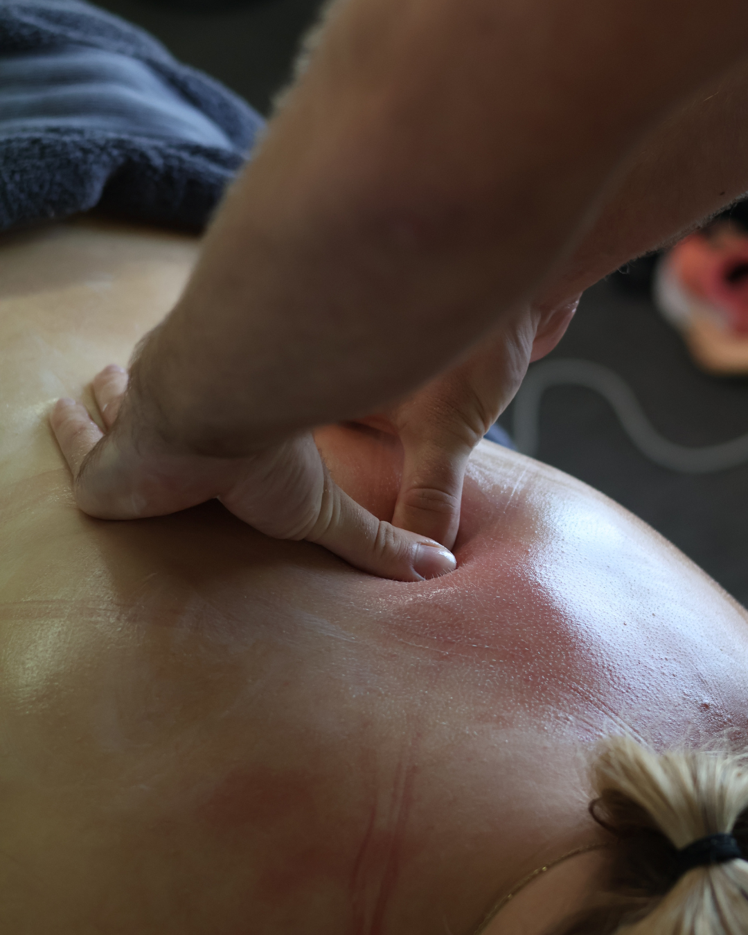
- Manual therapy
- Hands-on treatment — joint mobilisation of the glenohumeral and acromioclavicular joints, soft tissue therapy for the rotator cuff, posterior capsule and periscapular muscles, and dry needling for trigger points — reduces pain and improves movement quality in the short term. This creates the window needed to engage effectively with strengthening and movement retraining.
- Joint mobilisation of the cervical and thoracic spine is also valuable — the shoulder does not work in isolation from the neck and upper back, and restrictions in these regions directly affect shoulder movement patterns and symptom levels.
- Movement retraining and posture
- Rounded shoulders and forward head posture alter scapular position and rotator cuff length-tension relationships, contributing to the altered movement patterns that drive shoulder pain. Addressing habitual posture — at the desk, during sport, during sleep — is part of the treatment picture. Small changes in how you position your shoulder during daily activities can make a significant difference to symptom levels while rehabilitation progresses.
- Load management
- Understanding which activities are loading the shoulder beyond its current capacity and modifying them intelligently — reducing overhead volume temporarily, adjusting training, modifying workplace tasks — allows symptoms to settle while strength is being rebuilt. The goal is never to stop all activity, but to find a level that the shoulder can tolerate while rehabilitation progresses.
- Corticosteroid injection
- For presentations where pain is severe enough to prevent meaningful rehabilitation, a corticosteroid injection can provide short-term relief that creates the window for exercise to be effective. The evidence supports injection as an adjunct to physiotherapy rather than a standalone treatment — without the rehabilitation component, pain typically returns within months.
- Surgery
- Based on the current evidence, subacromial decompression surgery should not be a first-line treatment for rotator cuff related shoulder pain. A genuine trial of structured physiotherapy — not just a few generic exercises — is the appropriate first step for most presentations. Surgery may have a role in specific situations where conservative management has genuinely failed, but it is far less routinely indicated than it was a decade ago.
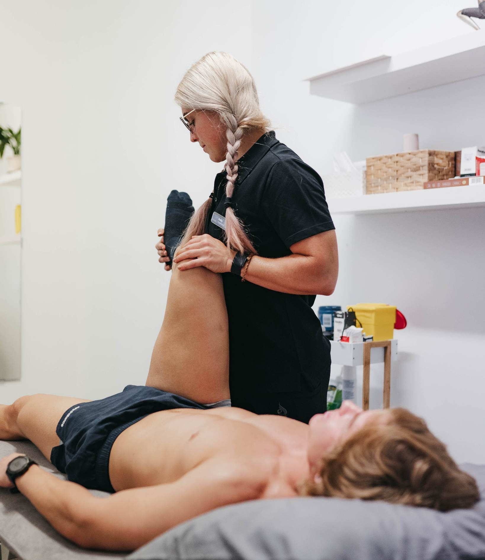
What to Expect at Active Balance
Our approach to shoulder pain starts with a thorough assessment of the entire shoulder complex — rotator cuff strength, scapular control, glenohumeral mobility, cervical and thoracic contribution, and the specific loading pattern driving your symptoms. This assessment drives a specific treatment plan rather than a generic shoulder program.
Treatment typically combines hands-on work to settle symptoms and restore movement with a progressive strengthening program built around your specific weakness pattern. The program is progressed systematically over weeks and months — meaningful rotator cuff and scapular strength improvements take time, and the most common reason shoulder pain recurs is stopping rehabilitation before adequate strength has been developed.
For people with shoulder pain related to sport — throwing, swimming, overhead lifting, racket sports — rehabilitation includes sport-specific loading and movement retraining to ensure the shoulder is genuinely ready for the demands being placed on it.
If shoulder pain is limiting your daily life or sport, book online or call us on (08) 7123 4148. We would love to help you understand what's actually driving your symptoms and get your shoulder moving comfortably again.